恒温器・振とう培養器等

取扱説明書の閲覧は学内からのみ行えます。

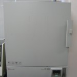
  • 恒温器 ヤマト科学 IC600
  • IC600_301_01
    設置場所: 203 試料調整室, 301共同利用実験室, 402 微生物培養室(2台)
    仕様: 150リットル、室温+5℃〜60℃
    取扱説明書(学内のみ)
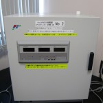
  • マルチガスインキュベータ アステック APMW-36
  • APMW-36_402_01
    設置場所:402 微生物培養室, 405 動物培養室
    仕様:CO2濃度範囲:0〜19.9%, O2濃度範囲:1.0〜94.0%, 室温+5℃〜50℃, ウォータージャケットタイプ, 内容量:36ℓ
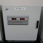
  • 炭酸ガスインキュベータ アステック APCW-36
  • APCW-36_01
    設置場所:405 動物培養室
    仕様:CO2濃度範囲:0〜19.9%, 室温+5℃〜50℃, ウォータージャケットタイプ, 内容量:36ℓ
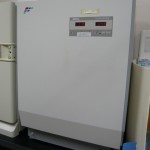
  • 炭酸ガスインキュベータ アステック SCI-165
  • SCI-165_01
    設置場所:405 動物培養室
    仕様:CO2濃度範囲:0〜19.9%, 室温+5℃〜50℃, ウォータージャケットタイプ, 内容量:163ℓ
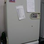
  • 炭酸ガスインキュベータ サンヨー MCO-17AI
  • MCO-17AI_01
    設置場所:405 動物培養室
    仕様:CO2濃度範囲:0〜20.0%, 室温+5℃〜50℃, ヒータジャケット+エアジャケット方式, 内容量:164ℓ
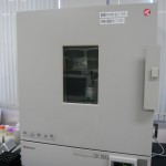
  • 乾熱滅菌器 ヤマト科学 DK600
  • DK600_203_01
    設置場所: 203 試料調整室
    仕様: 150リットル、40℃〜210℃
    取扱説明書(学内のみ)
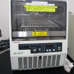
  • 恒温振とう培養器 タイテック BR-15
  • BR-15LF_01
    設置場所:301 共同利用実験室, 402 微生物培養室(2台)
    仕様:室温+5℃〜70℃, 旋回/往復振とう切換式 20〜200r/min
    取扱説明書(学内のみ)
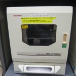
  • 恒温振とう培養器 タイテック BR-23FP
  • BR-23FP_301_01
    設置場所:301 共同利用実験室
    仕様:+15℃〜+60℃, 往復/旋回振とう切換式 20〜300r/min
    取扱説明書(学内のみ)

  • ブロック恒温槽 タイテック DTU-1B
  • 設置場所: 205 学生実験室, 301 共同利用実験室, 303 RNA実験室
    仕様:1.5mℓ用、室温+5℃〜110℃

  • 低温ブロック恒温槽 タイテック CTV-2515
  • 設置場所: 301 共同利用実験室, 303 RNA実験室
    仕様:1.5mℓ用、4℃〜50℃
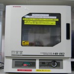
  • ハイブリダイゼーション・インキュベーター タイテック HB-80
  • HB-80_01
    設置場所: 301 共同利用実験室
    仕様:室温+5℃〜+80℃, 振とう速度 5〜60r/min, 振とう方式:シーソー、レシプロ、ボトル回転、チューブ転倒撹拌
    取扱説明書(学内のみ)

  • 小型回転培養機 ローテーター タイテック RT-5
  • 設置場所:203 試料調整室
    仕様:回転速度:0.5~5.0r/min
    取扱説明書(学内のみ)
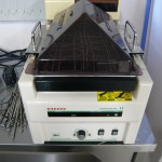
  • 振とう恒温槽(ウォーターバス) タイテック PERSONAL-11EX
  • Personal-11_01
    設置場所:205 学生実験室, 303 RNA実験室, 407 低温室
    仕様:往復振とう, 振とう速度:20〜160回/min, 振幅:10〜40mm可変式, 室温+5℃~+70℃
    取扱説明書(学内のみ)
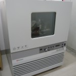
  • 振とう培養器 ヤマト科学 IM400W
  • IM400W_01
    設置場所:407 低温室(前室)
    仕様:5〜60℃, 水平回転振とう, 約30〜200回/min 70mm, 500mℓフラスコ×9個、1,000mℓフラスコ×5個
    取扱説明書(学内のみ)
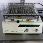
  • 振とう機 タイテック NR-3
  • NR-3_01
    設置場所:205 学生実験室, 301 共同利用実験室, 402 微生物培養室
    仕様:往復振とう, 振とう速度:20〜200回/min, 旋回/往復切換式
    取扱説明書(学内のみ)
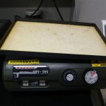
  • 撹拌振とう機 タイテック PR-36
  • PR-36_01
    設置場所:301 共同利用実験室
    仕様:波動形揺動, 20〜120回/min
    取扱説明書(学内のみ)

分光光度計(ナノドロップ ND-8000)取扱説明会

分光光度計(ナノドロップ)に関する取扱説明会の開催について

ナノドロップ(ND-8000)に関する取扱説明会についてお知らせします。
なお、今回は実機を用いて行いますので希望を募り、こちらで時間を調整致します。受講希望者は施設管理室までご連絡下さい。
*資料の関係上、9月2日(水曜)を期限とします。

  • 日時:2009年9月4日(金曜日)
    午前10時00分~午前10時40分 定員5名
    午前11時00分~午前11時40分 定員5名
    午前13時00分~午前13時40分 定員5名
    午後14時00分~午後14時40分 定員5名
    さらに人数が増える場合は午後の部を増やします。
  • 内容:
    1. ND8000について概要説明 (10分)
    2. 基本操作およびアプリケーションの説明 (25分)
    3. アプリケーションの紹介 (10分)
    4. 質疑応答 (5分)
  • 場所:遺伝子実験施設 試料調製室(2F)
  • 担当:エル・エム・エス 担当 黒瀬純志

第24回遺伝子実験施設セミナーの開催について

下記の要領で第24回遺伝子実験施設セミナーを開催します。
多数のご参加をお待ちしております。

  • 日時 8月18日 (火曜日) 午後3時から午後5時まで
  • 場所 遺伝子実験施設 1階 セミナー室
  • 講演 Special function unit in spermatic cord region
  • 講師 Dr. Worawut RERKAMNUAYCHOKE (タイ王立カセサート大学 獣医学部 准教授)
  • 概要

    Testicular artery and vein in the spermatic cord region play a major role in temperature regulation of testis through heat exchange mechanism.
    In addition, counter-current transfer of several kinds of substances,
    especially testosterone hormone, as well as gas between testicular artery and vein in the spermatic cord region has been demonstrated in several kinds of animals including man. Recently, using blood flow method showed blood bypassed the testis between 28 – 46%. Many suggestions were raised to explain this phenomenon, such as, the mechanism may be accomplished by arterio-venous anastomosis in the cord or it may be done by diffusion. In order to fulfill the requirements of the target organs (testis or epididymis), it is considered that the structure of testicular artery and vein in the cord has been modified. However, the morphological architecture which regards to this evidence in the cord has not been fully understood.

    The aims of these studies were designed to evaluate any kind of special function unit that presents in spermatic cord region. Six species of animals, golden hamster, swine, common tree shrew, variable squirrel squirrel, Asian musk shrews and lesser mouse deer, were studied by light and transmission electron microscopies as well as corrosion cast techniques combined with scanning electron microscopy.

    The special function unit was found in spermatic cord region in these studies and could be classified as primary and secondary modifications. The primary modifications were shared characters such as convoluted testicular artery, thinner vascular walls between testicular artery and apposition vein as well as anastomosis between veins. While, the secondary modifications were considered to be species – dependent characters. This secondary modifications contained with vesiculated cells and direct arterio-venous anastomosis between testicular artery and vein in common tree shrew and variable squirrel, vesiculated capillary complex in lesser mouse deer, venous portal system in swine, endothelial bridge of the venous walls as well as abundance mast cells in the base of the protruding venous wall in golden hamster and extratesticular Leydig cell in Asian musk shrew.